Bone Profile
Bone profile examines the proteins, minerals and enzymes involved in bone metabolism. Discrepancies with these can indicate a problem with bone turnover. Bone profile typically tests the following. Normal reference values in brackets.
- Serum Total Protein (64.00 – 83.00 g/L)
- Serum Albumin (35.00 – 50.00 g/L)
- Serum Calcium (2.10 – 2.55 mmol/L)
- Serum Inorganic Phosphate (0.80 – 1.50 mmol/L)
- Corrected Serum Calcium level (2.10 – 2.55 mmol/L)
- Serum Globulin (20.00 – 39.00 g/L)
- Serum Alkaline Phosphatase (40.00 – 150.00 U/L)
- Protein – Low total protein levels can suggest a liver (albumin is produced in liver. All major blood proteins apart from immunoglobulins are synthesised in liver) or kidney disorder (in nephropathy, is excreted from kidney), or a disorder in which protein is not digested or absorbed properly, or immunoglobulin not being made (for example in bone marrow failure) High total protein levels can indicate dehydration, or some types of cancer, that lead to an accumulation of an abnormal protein (such as immunoglobulin in multiple myeloma)
- Calcium – can be high in blood – hypercalcemia – in certain bone cancers and hyperparathyroidism.
- Alkaline phosphatase (ALP) – is an enzyme which is found in high amounts in bone and liver. Raised ALP indicates a problem with bone (raised in Pagets, Osteomalacia, certain cancers) or liver. If calcium and phosphate are also altered, then it is related to bone metabolism but if bilirubin, GGT and ALT are raised then it is coming from the liver.
Myeloma Screen
Multiple Myeloma is a primary blood cancer which affects plasma cells. Most of these plasma cells are present in bone marrow. As it takes up space in the bone marrow it leaves less room for the normal cells thus causing anemia, leukopenia and thrombocytopenia.
Plasma cells are immune systems cells that are involved in making antibodies. Myeloma cells also make antibodies called monoclonal antibodies which do not fight infection. The monoclonal antibodies are made up of two types of proteins. Bence Jones protein is one of them. These show up in urine of people with multiple myeloma.
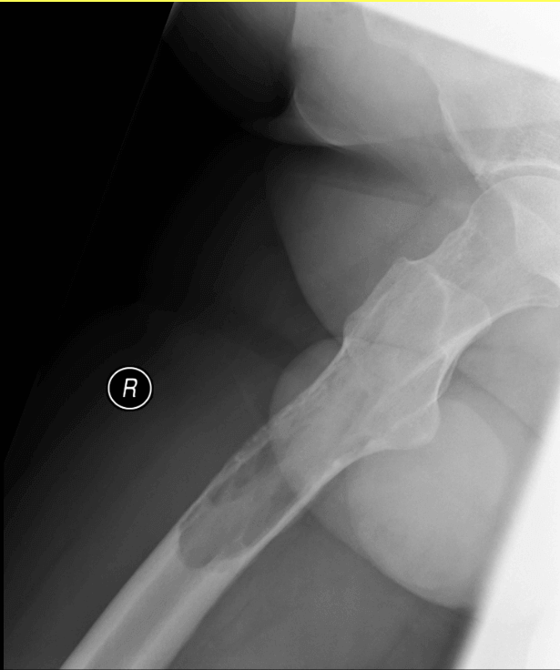
As myeloma cells activate osteoclasts, they start to breakdown bone and weaken them which can cause fractures but also leads to higher calcium levels in the blood (hypercalcemia).
- FBC
- ESR, CRP
- Urea & Electrolytes
- Calcium, Phosphate, Albumin
- Serum & Urine Electrophoresis – electrophoresis in general means the separation of charged particles under the influence of an electric field.
- Serum Electrophoresis separates proteins in a blood sample based on their charge when an electric current is applied for a predetermined time. Mainly Albumin and Globulin (alpha-1, 2 globulins, beta globulins and gamma globulins) bands are seen. These are divided in to 5 major fractions (Albumin, Alpha-1 zone, Alpha-2 zone, Beta zone & Gamma zone). In Multiple Myeloma an abnormal “M Band” appears on the electrophoresis strip. This is also called a paraprotein. This indicates monoclonal gammopathy. Most commonly observed paraprotein is IgG (70% of times) followed by IgA (20% patients), light chain (5-10%) and IgD, E or M (very rare, <1%). The identification of exact type of paraprotein requires ‘immunofixation’.
- Urine Electrophoresis is done to detect light chain monoclonal paraprotein called Bence-Jones Protein in the urine. These are mainly present in Multiple Myeloma or Lymphoma.
Why does ESR get raised in Multiple Myeloma
2 answers on quora are from internal medicine doctors.
Acute phase reactant proteins (fibrinogen in particular) cause erythrocytes to settle to the bottom of a test tube at a faster rate. Acute phase reactants are proteins that increase in concentration in the blood in states of chronic inflammation. Inflammation is prevalent in various types of cancer.
Multiple myeloma is a form of cancer involving the plasma cells of the bone marrow. Thus, chronic inflammation associated with it is one explanation for the increased ESR in multiple myeloma. In addition, various immunoglobulins, particularly IgG, IgA and IgM can raise the sedimentation rate. Since the plasma cells of multiple myeloma can produce an increased number of either or a combination of these proteins, this is another explanation for the increased ESR of multiple myeloma.
Victor Battles, M.D Internal Medicine, Perelman School of Medicine at the University of Pennsylvania (1978)
Multiple myeloma is a malignant disease where the tissue type involved is b-cells. b-cells generally produce antibodies and in multiple myeloma increased levels of abnormal antibodies are the usual. These antibodies can coat red blood cells and interfere with the normal electrical charge on the red cell membranes that causes red cells to repel each other. Thus, the sedimentation rate is increased.
Tony Jay, former ICU Physician